Objective Lenses for Liquid
Crystal Observation
Both Nikon and Evident
(formerly Olympus) offer objective lenses specifically designed for
liquid crystal display (LCD) cell observation, as indicated by their
model names. However, it’s rare to see these lenses being used by
liquid crystal researchers. This raises questions about where these
lenses are actually used and whether they offer any advantages for
liquid crystal research. I will explore these questions and offer
some speculative insights.
Correction Collar Objectives
Microscope objective lenses are typically corrected for
specific observation conditions. For example, most biological
objectives are optimized for viewing through a cover glass with a
standard thickness of 0.17 mm, while metallurgical objectives are
designed for observation without a cover glass. Deviation from these
optimized conditions can lead to spherical aberrations, causing
image blurring and reducing effective resolution. This effect is
particularly pronounced with high numerical aperture (NA)
objectives, where even a 0.01 mm deviation in cover glass thickness
can result in noticeable image degradation.
Correction collar
objectives, which include the liquid crystal cell objectives, allow
for adjustments to minimize image degradation when observing through
glass plates of varying thicknesses. These objectives feature a
rotating collar with a scale on the body of the lens, allowing for
adjustment based on the thickness of the glass. Correction collar
objectives can be broadly categorized into three types:
1. High NA, High
Magnification Apochromat or Semi-Apochromat Lenses;
These are designed to correct for variations in cover glass
thickness, typically within the range of 0.11–0.22 mm.
2. Long Working Distance
Objectives for Inverted Microscopes; These
are often used for observing samples in petri dishes, with a
correction range that can accommodate glass thicknesses from 0 to 2
mm.
3. Specialized Objectives
for Specific Applications; These
include lenses designed for observing CDs or optical discs, as well
as liquid crystal cells.
Usage of Liquid Crystal
Correction Collar Objectives
Liquid crystal cell
objectives seem to be used primarily in manufacturing settings, such
as for inspecting LCD panels. However, it’s unlikely that these
lenses are being used to observe the liquid crystals themselves, as
the liquid crystals in such panels are typically well-aligned and
lack dramatic defect structures. Instead, these objectives are
probably used to inspect the electrodes and transistors within the
cells. An engineer I spoke with mentioned that, in some cases, the
correction collar was set incorrectly, yet adjustments were not made
because the inspection criteria had already been established based
on the incorrect setting. This suggests that correction collar
objectives are not always used correctly, even in industrial
settings.
Setting the Correction Collar
One reason for the incorrect
use of correction collars may be that users lack an understanding of
what constitutes an optimal image. Since it’s difficult to describe
in words, I will illustrate the difference with examples.
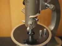
I used two finite conjugate
system long-working-distance objectives—a 40x NA 0.5 metallurgical
objective and a 40x NA 0.55 biological objective—for comparison.
Although I own liquid crystal observation objectives, I do not have
a matching infinity-corrected microscope, so this discussion will be
based on finite systems. Metallurgical long-working-distance
objectives are also available for infinity systems and are commonly
used with hot stages. However, biological long-working-distance
objectives often lack sufficient working distance for use with
commercially available hot stages, necessitating custom hot stage
setups.
The sample used for this
comparison is a diatom slide from MicroWorld Services. For
resolution testing, the test plate or the J series for viewing
should be used, as the diatoms are in close contact with the cover
glass, which is of standard thickness. This ensures that the best
image achievable with the objective lens is automatically obtained,
even if the user does not know what the best condition is.
First, I will show an image
of the diatoms observed through the cover glass using the biological
objective. This objective allows for adjustment to match the cover
glass thickness of 0.17 mm.
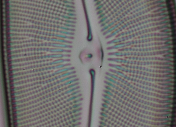
Fig.
Observation through a
cover glass using the biological objective.
Next, I will show an image taken through the cover glass using
the metallurgical objective.
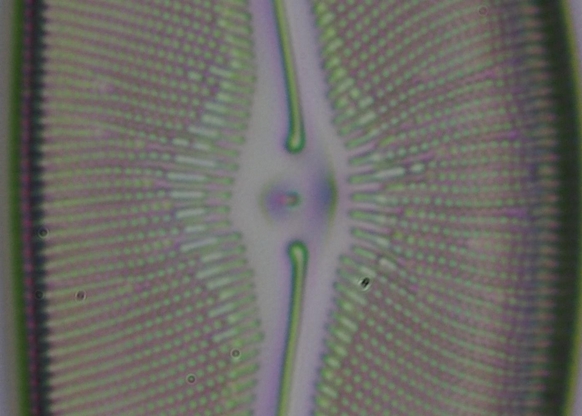
Fig. Observation through the cover glass using the
metallurgical objective.
The metallurgical objective is designed for observation without a
cover glass, so using it with a cover glass introduces spherical
aberrations, leading to blurred images. Compared to the biological
objective, the lines appear broader and the contrast between
adjacent points is weaker.
Next, the diatom slide was flipped, and observations were made
through the slide glass, which is about 1 mm thick. This deviates
even further from the objective lens's designed conditions. This
setup simulates conditions similar to those in liquid crystal
research, where samples are often observed through thick slide glass
or ITO glass, sometimes through a hot stage window.
I will show two images taken with the metallurgical objective,
with focus positions adjusted.
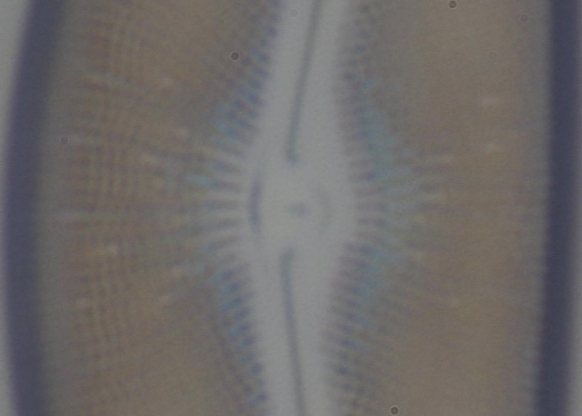
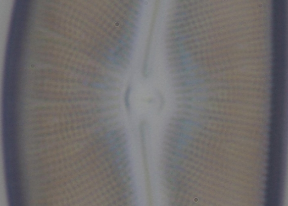
Fig.: Observations through the slide glass using the metallurgical
objective (two focus positions).
Compared to the cover glass observations, there is significant image
degradation due to the increased blurring. The lower image shows
better resolution of the dots on the left and right sides, while the
upper image has better contrast and clarity in the central vertical
line. However, these differences should not be interpreted as
differences in depth within the sample. Instead, they are artifacts
of spherical aberrations caused by the misalignment of the imaging
positions of low NA (contributing to coarse structures) and high NA
light (contributing to fine structures).
Finally, I will show an image taken through the slide glass using
the biological objective, properly adjusted using the correction
collar.
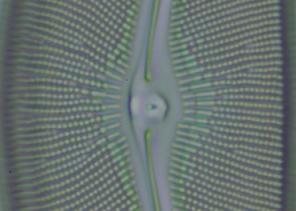
Fig. Observation through the slide glass using the biological
objective with proper correction.
Unlike the metallurgical objective, this image shows little
difference compared to the cover glass observation, demonstrating
the effectiveness of the correction collar. Proper adjustment of the
collar is crucial; otherwise, the image will be blurred.
In
my experience, correction collar objectives are rarely used in
liquid crystal research. This is partly due to the lack of
correction collar objectives with sufficient working distance for
use with hot stages. Additionally, liquid crystal structures
generally have high contrast, and fine structure is often less
critical, reducing the perceived need for such objectives. However,
for researchers who want to improve the observation of fine
structures, correction collar objectives offer a valuable tool.
|